微信公众号
微信公众号

 投诉邮箱
投诉邮箱
 监督电话
监督电话

最新消息


【公共营养与健康食品产业】践行健康中国战略,促进农业与食品和大健康产业发展的公共服务与宣传。在中食协营养委专家智库指导下,共同关注国民营养与健康;关注科技创新与可持续发展;关注食品安全和农业与食品全产业链发展;关注政策法规行业标准;关注食育科普、健康管理、营养配餐;关注特医食品、药食同源、功能食品开发应用;关注产学研一体化;关注地方特色产业集聚开发利用。
本平台由中国食协营养委员会监督指导,北京佳源汇咨询有限公司维护运营。


-

第七届(IFSN)国际会议
“第七届(IFSN)国际营养健康与食品安全峰会”暨“第十三届中国食药同源功能创新产业大会”定于2025年9月21-24日在北京召开。
2025-05-27 第七届(IFSN)国际会议 -

-

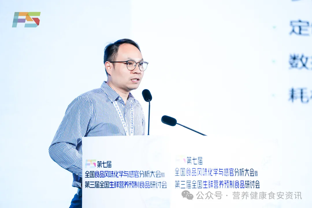
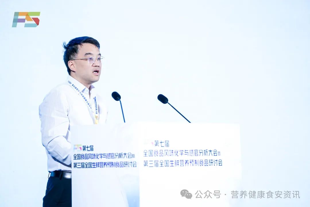
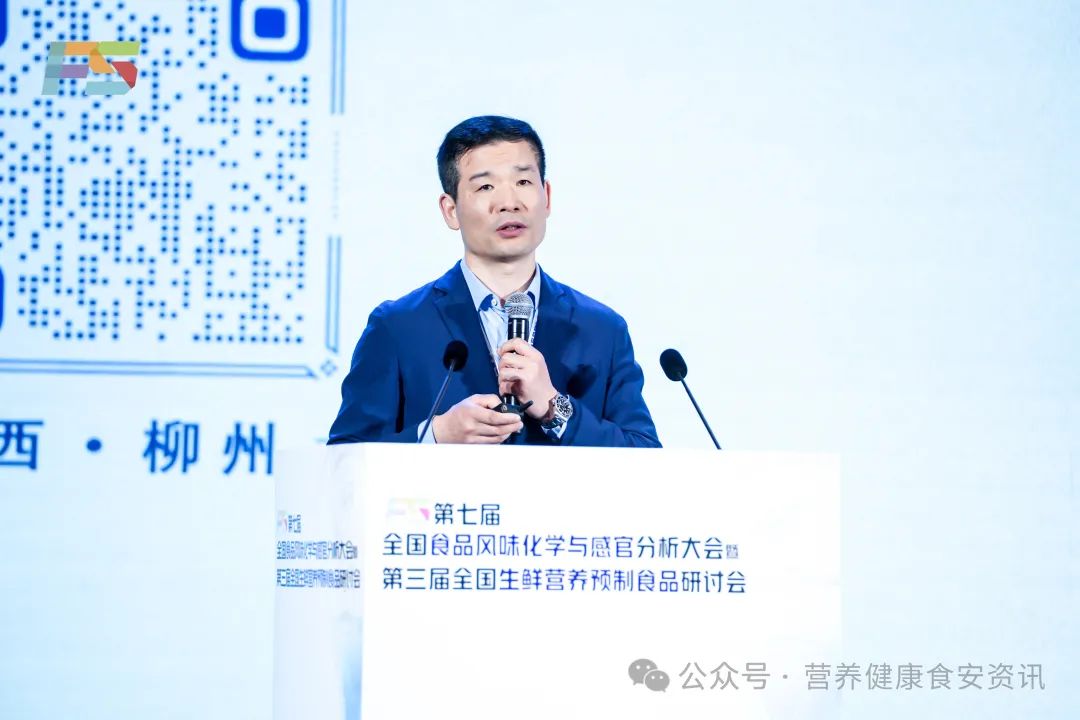
“第七届全国食品风味化学与感官分析大会” 论文、墙报展示、青年论坛发言申请表
欢迎下载“第七届全国食品风味化学与感官分析大会”论文、墙报展示、青年论坛发言申请表。
2024-12-26 “第七届全国食品风味化学与感官分析大会” 论文、墙报展示、青年论坛发言申请表 -
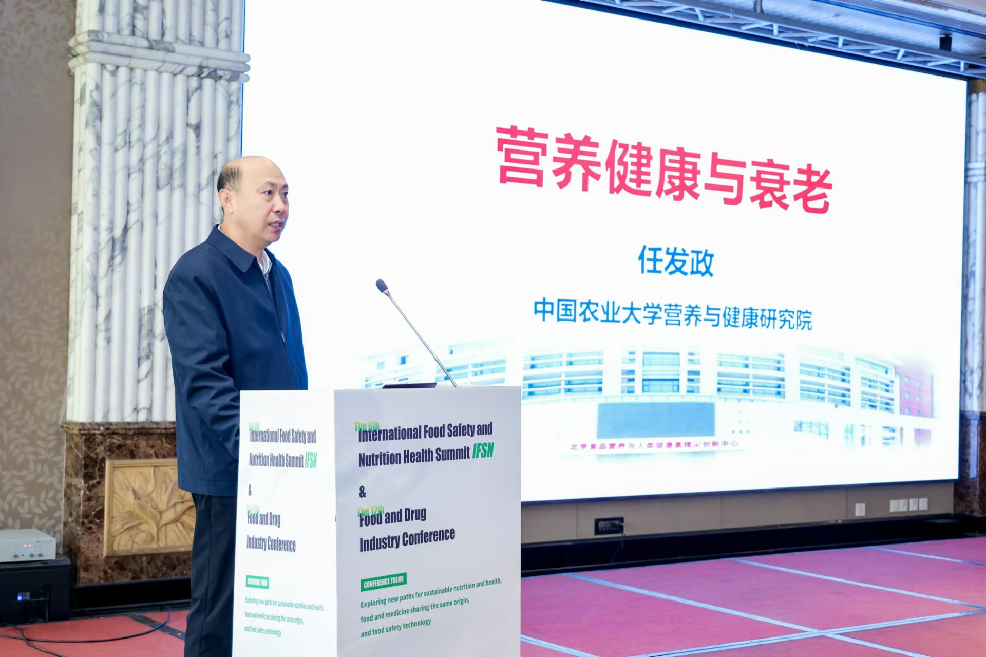
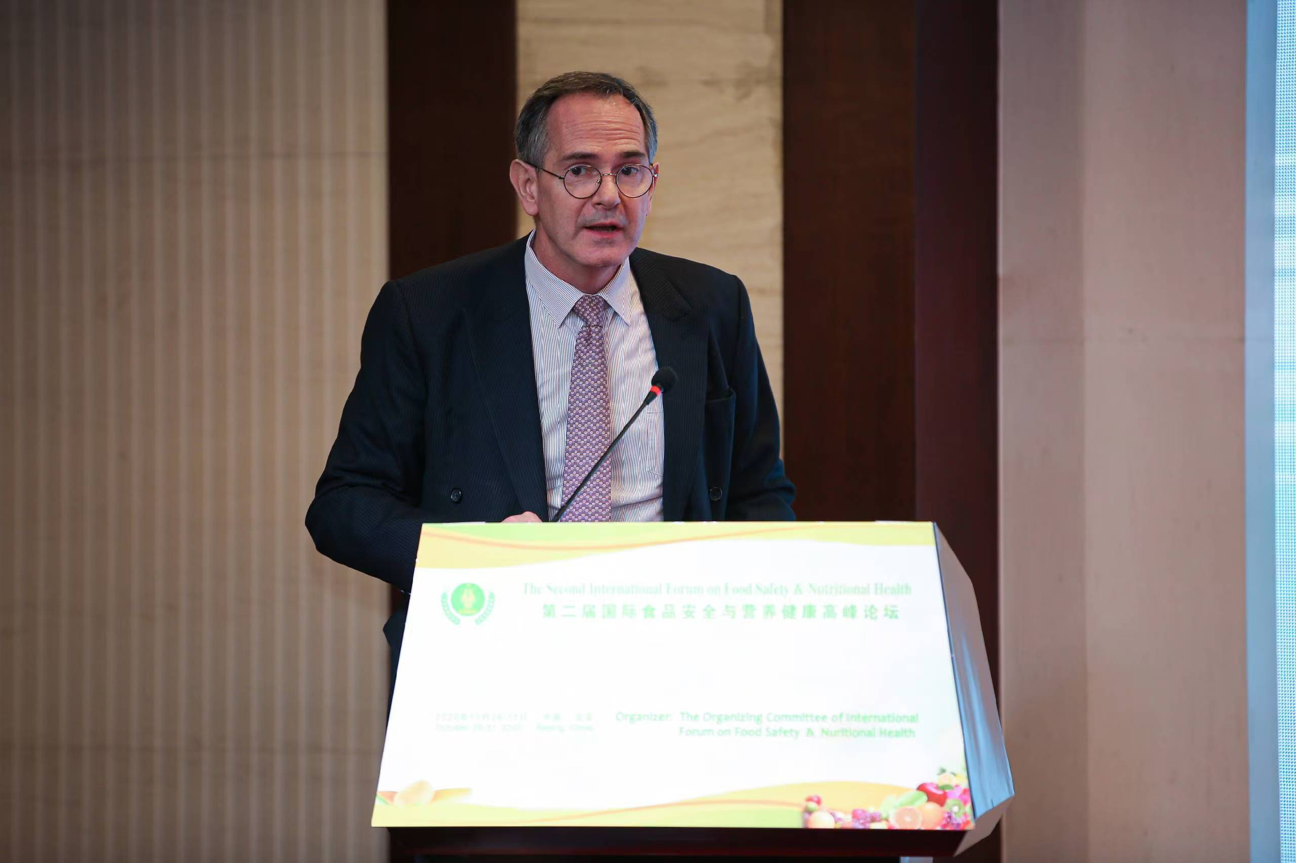
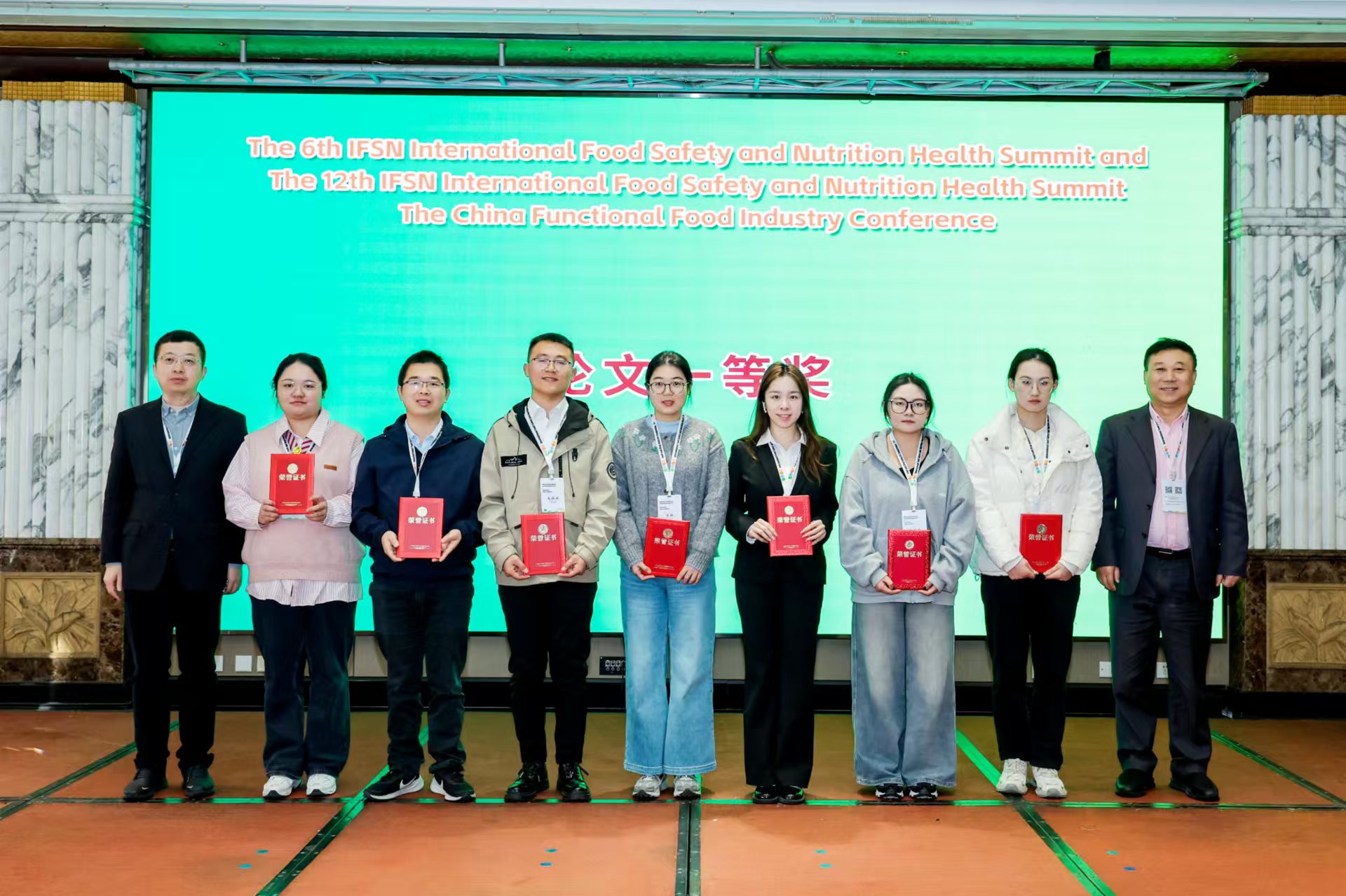
“第六届(IFSN)国际食品安全与营养健康峰会”暨“第十二届中国功能食品产业大会”在京召开
为培育并激活食品产业新质生产力,扩大营养健康安全食品科技赋能应用场景,以“探索可持续营养健康、药食同源、食品安全科技新路径”为主题备受欢迎的,“第六届(IFSN)国际食品安全与营养健康峰会”暨“第十二届中国功能食品产业大会”近日在北京召开.
2024-11-22 “第六届(IFSN)国际食品安全与营养健康峰会”暨“第十二届中国功能食品产业大会”在京召开 -

更多内容
-

关于组织开展“健康中国·营养创新万里行” 考察调研活动的通知
将科研创新与产业升级、企业实践有效紧密结合,以“科技”加持带动营养健康产业、地方经济发展,我会决定组织“健康中国·营养创新万里行”考察活动。
2025-06-06 关于组织开展“健康中国·营养创新万里行” 考察调研活动的通知 -
-
-
-

更多内容
-
-
-
-
-

田明研究员在“第六届(IFSN)国际食品安全与营养健康高峰论坛”暨“第十二届中国功能食品产业大会”上的演讲
功能性食品的定位及科学构建食品声称管理体系的思考与建议
2024-12-09 田明研究员在“第六届(IFSN)国际食品安全与营养健康高峰论坛”暨“第十二届中国功能食品产业大会”上的演讲
更多内容
-

中共中央办公厅 国务院办公厅关于进一步强化食品安全全链条监管的意见
为进一步理清食品安全监管责任,建立健全协同监管机制,强化全链条监管合力,坚决守牢食品安全底线,切实保障人民群众身体健康和生命安全.
2025-07-02 中共中央办公厅 国务院办公厅关于进一步强化食品安全全链条监管的意见 -
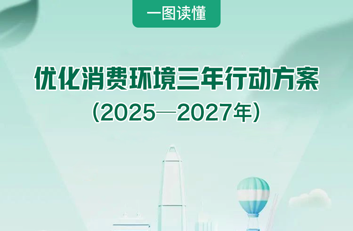
市场监管总局等五部门印发《优化消费环境三年行动方案(2025—2027年)》(附一图读懂)
优化消费环境是提振消费信心、激发经济活力的重要举措,对推动经济高质量发展、保障高品质生活具有重要意义。
2025-06-17 市场监管总局等五部门印发《优化消费环境三年行动方案(2025—2027年)》(附一图读懂) -

《2025项目指南-卓越研究群体项目》--国家自然科学基金委员会
致力科学前沿突破,产出一批国际领先水平的原创成果,抢占国际科学发展的制高点,形成若干具有重要国际影响的学术高地。
2025-05-23 《2025项目指南-卓越研究群体项目》--国家自然科学基金委员会 -
《2025项目指南-国家杰出青年科学基金项目》--国家自然科学基金委员会
旨在促进青年科学技术人才的成长,吸引海外人才,培养和造就一批进入世界科技前沿的优秀学术带头人。
2025-05-23 《2025项目指南-国家杰出青年科学基金项目》--国家自然科学基金委员会 -
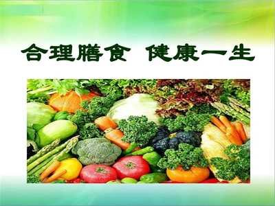
国民营养健康指导委员会办公室关于印发“健康饮食、合理膳食”核心信息的通知
2025年围绕“健康饮食、合理膳食”主题,倡导增加蔬菜水果、全谷物和水产品摄入,加大针对性科普宣传,引导形成合理的膳食结构。
2025-05-22 国民营养健康指导委员会办公室关于印发“健康饮食、合理膳食”核心信息的通知
更多内容
-
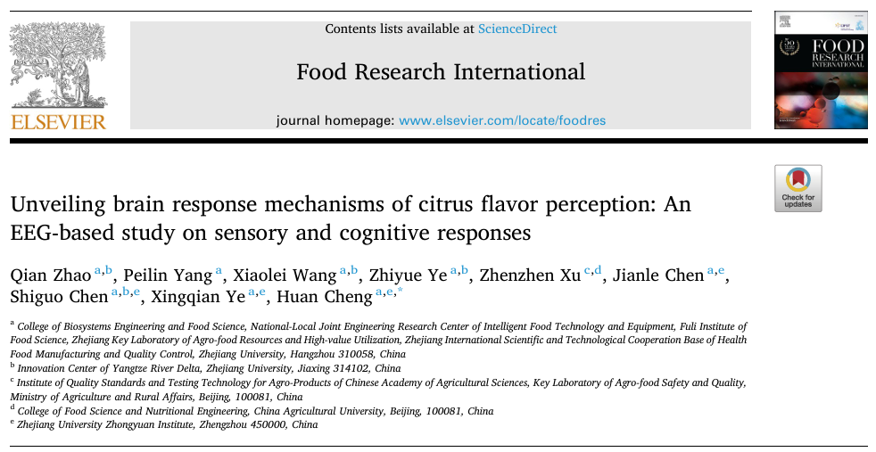
揭示柑橘风味感知的大脑反应机制:一项基于脑电图的感觉和认知反应研究
这是首个基于脑电图的研究,用于比较大脑对不同柑橘香味的反应,为食品行业在优化产品配方和增强消费者体验方面提供了重要启示。
2025-07-01 揭示柑橘风味感知的大脑反应机制:一项基于脑电图的感觉和认知反应研究 -
-
豌豆蛋白与三种含硫风味化合物的相互作用:分子结构、非共价和结合机制的见解
本研究通过多光谱光谱技术和分子对接,首次系统性地探究了含硫风味化合物(SCFCS)与豌豆蛋白分离物(PPI)之间的相互作用机制。
2025-06-24 豌豆蛋白与三种含硫风味化合物的相互作用:分子结构、非共价和结合机制的见解 -
大豆分离蛋白和植物肉在储存过程中挥发性异味化合物的形成
本研究调查了导致挥发性化合物发展的因素以及导致异味的挥发性化合物。本研究的主要目的是研究大豆分离蛋白和植物肉在贮藏过程中挥发性异味化合物的形成。
2025-06-18 大豆分离蛋白和植物肉在储存过程中挥发性异味化合物的形成 -
海参蛋白与风味物质的相互作用机制:吸附行为、动态平衡和结构变化 风味化学感官分析 食品风味化学与感官分析 2025年06月14日 20:00 云南
本研究通过分析 CCE 的构象变化和这些风味物质的吸附行为,为理解海参蛋白与风味物质之间的相互作用机制奠定了基础。
2025-06-16 海参蛋白与风味物质的相互作用机制:吸附行为、动态平衡和结构变化 风味化学感官分析 食品风味化学与感官分析 2025年06月14日 20:00 云南
-

我国国际食品科学院院士汇总(二十一)--江南大学食品学院 --江 波院士
最先研究低聚果糖生产的研究者之一,并作为学术带头人率先实现了低聚果糖在国内的工业化生产。他在国内率先从事研究的成果还包括谷氨酰胺转胺酶、伽玛氨基丁酸、双果糖苷、塔格糖、阿洛酮糖和非还原性糊精等生物或功能食品配料。
2025-05-29 我国国际食品科学院院士汇总(二十一)--江南大学食品学院 --江 波院士 -

我国国际食品科学院院士汇总(二十)--北京三元食品股份有限公司 -- 陈历俊院士
助力中国母婴营养健康水平提升,科技引领中国奶业持续健康发展。
2025-05-29 我国国际食品科学院院士汇总(二十)--北京三元食品股份有限公司 -- 陈历俊院士 -

我国国际食品科学院院士汇总(十九)--浙江省农业科学院食品科学研究所 --郜海燕院士
主要从事食品物流保鲜加工与品质控制、生鲜食品未来采后与营养健康、植物源食品功能挖掘与综合利用等相关研究。
2025-05-29 我国国际食品科学院院士汇总(十九)--浙江省农业科学院食品科学研究所 --郜海燕院士 -
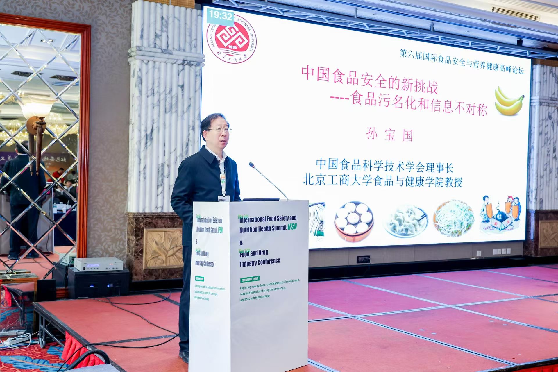
我国国际食品科学院院士汇总(十八)--北京工商大学 --孙宝国院士
构建了肉香味含硫化合物分子特征结构单元模型,研究成功了一系列重要肉香味食品香料制造技术,奠定了我国3-呋喃硫化物系列和不对称二硫醚类食品香料制造的技术基础
2025-05-26 我国国际食品科学院院士汇总(十八)--北京工商大学 --孙宝国院士 -
我国国际食品科学院院士汇总(十七)--中国农业科学院农产品加工研究所 --王 强院士
在国际上首次提出高水分挤压过程中蛋白纤维结构形成的“分层叠变”新理论,创建了基于高水分挤压的植物基肉制品颠覆性加工新技术,革新设计了智能化加工装备和可视化平台,引领了植物基肉制品产业发展新方向。
2025-05-26 我国国际食品科学院院士汇总(十七)--中国农业科学院农产品加工研究所 --王 强院士
更多内容


-


安捷伦:新型7010D三重四极杆气质联用系统
-

岛津水墨动画成片
-

东阿阿胶:大众最信赖的滋补健康引领者
-

稼速:链接全球好物,稼速新质生活
-

盈盛恒泰:提供专业的智能感官分析仪器
-

安琪酵母 创新健康生活
-

中科爱伽胖胖博物馆
-

滔普实验室:品质成就价值
-
 玛哈·艾哈迈德:联合国世界粮食计划署中国办公室副主任
-
 柯东凯:澳大利亚农业部北京驻华使馆农业参赞
-
 张春晖 :中国农科院农产品加工所中式食品加工与装备创新团队首席科学家
-
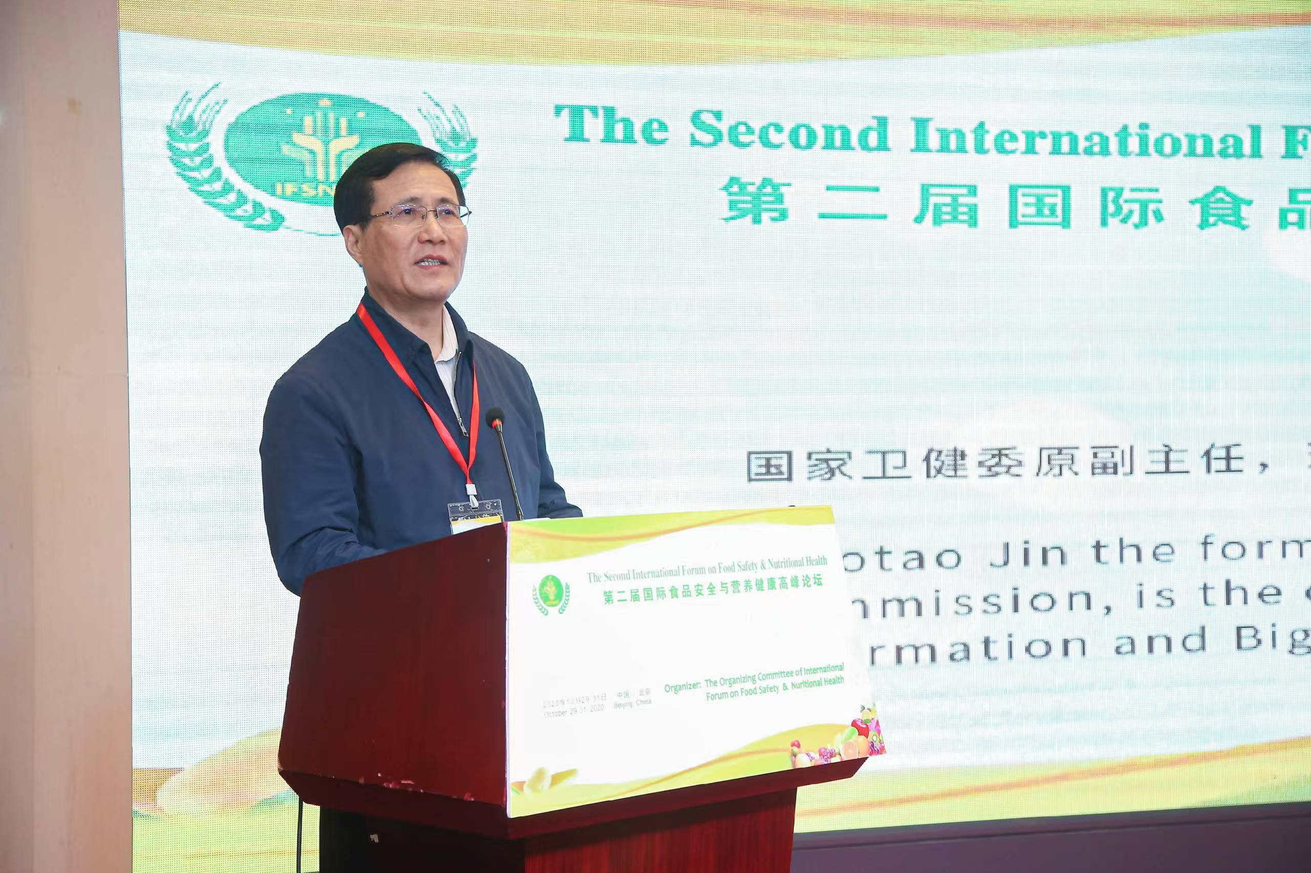 金小桃:中国卫生信息与健康医疗大数据学会会长
-
 毕玉安:国家食品药品稽查专员
-
 欧敏行:联合国粮食及农业组织驻华代表处 代理代表
-
 罗云波:国际食品科学院院士 北京工商大学食品与健康学院教授
-
 丁钢强:中国疾控中心营养与健康所所长 主任医师
-
 王兴国:江南大学食品学院教授
-
 赵谋明:华南理工大学食品科学与工程学院教授
-
 陈建设:国际食品科学院院士 原浙江工商大学特聘教授
-
 杜荷会长
杜荷会长
-
 刘秀梅:国际食品科学院院士 原中国疾控中心食品安全首席专家
-
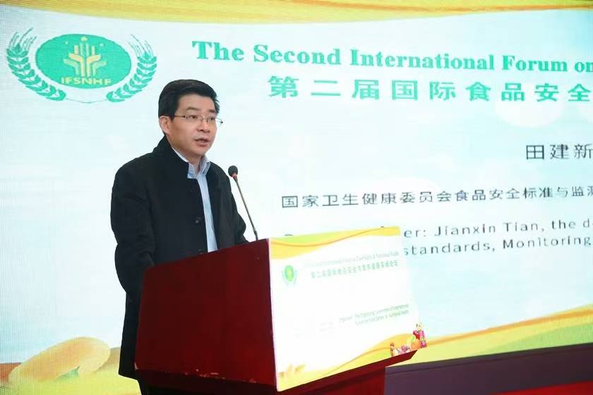 田建新:国家卫生健康委员会食品安全标准与监测评估司副司长